
Liquid Flow

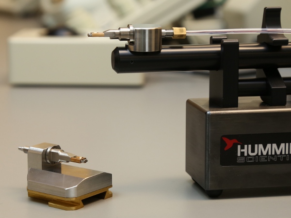
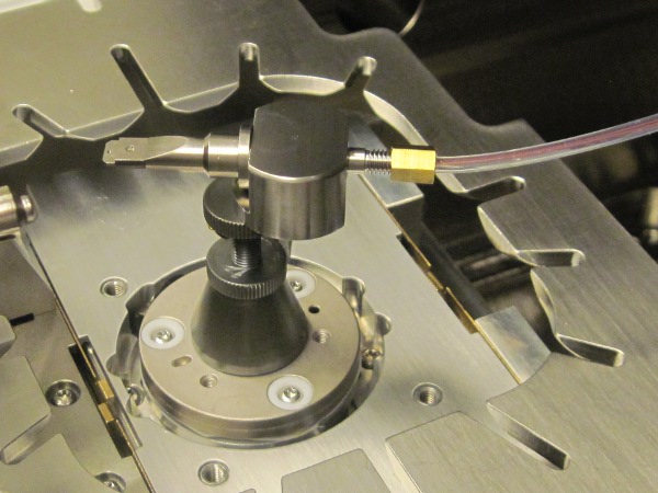
| 1400 Series SEM | |
| Number of Inlets | 1 or 2 depending on model, single outlet |
| Biasing Contacts | 4 |
| Tubing Type | User-replaceable microfluidic tubing |
| Delivery System | Variable-speed liquid delivery system |
| Tip Type | Removable tip |
| Flow Type | Continuous or static liquid flow |
| SEM Compatibility | Custom integration* |
Cross-Correlative Studies
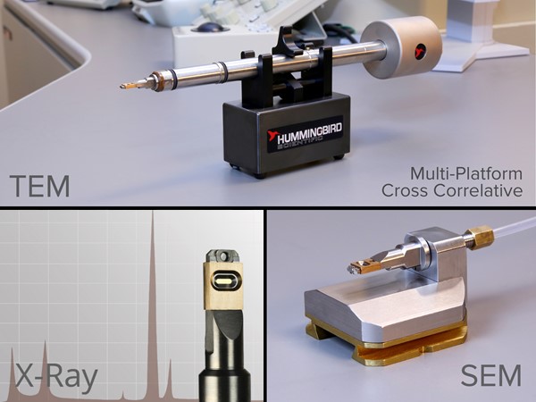
Researchers studying the interaction of biological structures with the environment have long been hindered by the lack of a single microscopy technique capable of spanning all relevant length scales. In response to this problem, we have developed systems that allow multiple correlative imaging techniques. The SEM liquid holder enables researchers to perform cross-correlative experiments and to image specimens in liquid environment. Using this system, researchers at Evergreen State College, Ames Laboratory, and the University of Georgia have successfully taken correlative images of biological specimens using LM, SEM, and TEM.
Figure: Correlating fluorescence (top) and SEM images of autofluorescent Anthrospira in phosphate-buffered saline (bottom), both taken using Hummingbird’s correlative liquid cell system.
Reference: D.A. Fischer, D.H. Alsem, B. Simon, T. Prozorov and N. Salmon. “Development of an Integrated Platform for Cross-Correlative Imaging of Biological Specimens in Liquid using Light and Electron Microscopies.” Microscopy and Microanalysis 19:Suppl. 2 (2013) pp. 476–477. Abstract
Edit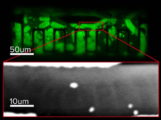
| Alejandra Londono-Calderon, Srikanth Nayaka, Curtis L. Mosher, Surya K. Mallapragadaa, Tanya Prozorov. “New approach to electron microscopy imaging of gel nanocomposites in situ.” Micron (2019) | Abstract |
| Alejandra Londono-Calderon, Wan Sun, Zengyi Shao, Daan Hein Alsem, and Tanya Prozorov. “Correlative Microbially-Assisted Imaging of Cellulose Deconstruction with Electron Microscopy.” Microscopy & Microanalysis (2018) | Abstract |
| D.A. Fischer, D.H. Alsem, B. Simon, T. Prozorov, and N. Salmon. “Development of an Integrated Platform for Cross-Correlative Imaging of Biological Specimens in Liquid using Light and Electron Microscopies.” Microscopy & Microanalysis (2013) | Abstract |
Read More

