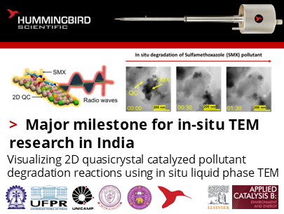

🎉 Proud to share a major milestone for in-situ TEM research in India! 🎉
We are delighted to congratulate Dr. Chandra Sekhar Tiwary, Dr. Shamik Chowdhury, and their outstanding team of collaborators from IIT Kharagpur, Federal University of Paraná (UFPR), State University of Campinas (UNICAMP), Indian Institute of Technology (BHU), and University of Delhi, who worked closely with Prikshat Dadhwal and Saka Pranith Chander from Hummingbird Scientific, India for their newly accepted paper:
“Radio Frequency-Induced Catalysis using Multi-Component Two-Dimensional Quasicrystals for Effective Sulfamethoxazole Removal from Water.”
This milestone marks the first peer-reviewed publication from India using a Hummingbird Scientific Gen IV liquid flow TEM sample holder, and we are proud to have supported this achievement.
The researchers developed a novel strategy for degrading the antibiotic sulfamethoxazole (SMX) using radio-frequency–activated two-dimensional AlFeCoNiCu quasicrystals (QCs). Their experiments revealed strong catalytic interactions between SMX molecules and the QC surface, supported by RF-catalysis measurements, spectroscopy, and computational modeling.
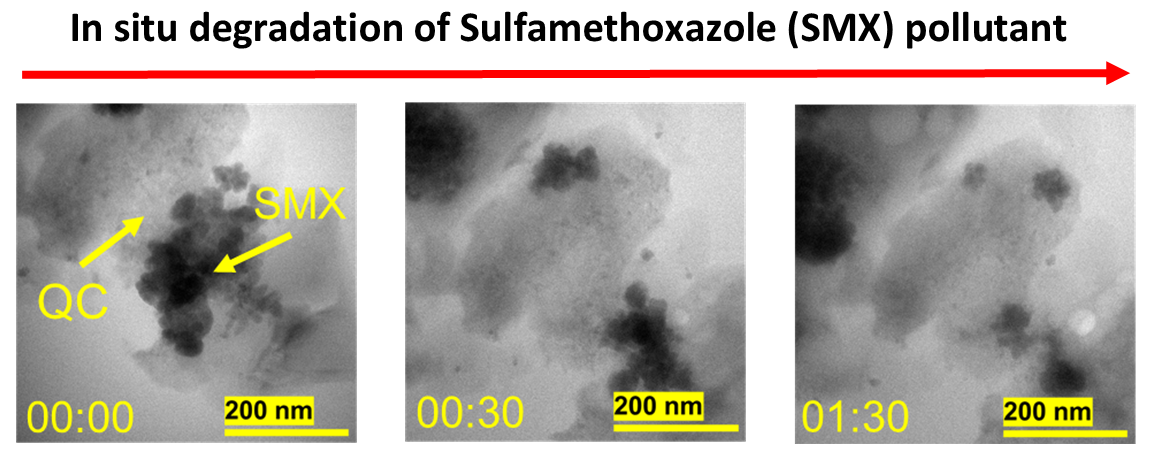
In situ liquid flow TEM images showing how sulfamethoxazole (SMX) molecules interact with and break down on the surface of 2D AlFeCoNiCu quasicrystals (QCs). At the start of the experiment (00:00), SMX appears as dark regions adsorbed onto the lighter QC surface. As time progresses (00:30 and 01:30), these dark areas gradually disappear, revealing the step-by-step degradation of SMX.
Using the Hummingbird Scientific Gen IV liquid flow TEM sample holder, the team directly visualized how 2D AlFeCoNiCu quasicrystals (QCs) interact with and progressively degrade the antibiotic sulfamethoxazole (SMX) in solution. The in situ holder made it possible to observe the entire degradation sequence in real time—something that cannot be achieved with conventional TEM or ex-situ characterization. By maintaining a stable, well-controlled liquid environment at high spatial resolution, the liquid cell enabled the researchers to capture rare nanoscale evidence of the process, beginning with the adsorption of SMX molecules onto the QC surface, visible as distinct dark contrast regions. As imaging continued, these regions gradually diminished, revealing the step-by-step breakdown and disappearance of SMX at the interface.
This publication is an exciting first step toward expanding advanced in-situ microscopy capabilities across India. We look forward to supporting more research that uses our in-situ platforms to study materials behavior, catalytic processes, electrochemistry, environmental remediation, and more. As more researchers gain access to these tools, we see India growing into a major center for innovative operando science—where real-time observation leads to new discoveries, new materials, and new technologies. Our goal is to help build a strong and collaborative scientific community, and this achievement shows what is possible when innovation and partnership come together.
Reference: Zahoor Manzoor, et. al., Appl. Catal., B Environ., 383, 126062 (2025) DOI: 10.1016/j.apcatb.2025.126062
Full paper Copyright © 2025 Elsevier B.V. All rights are reserved, including those for text and data mining, AI training, and similar technologies.
View All News