What does in-situ liquid-phase TEM reveal about crystallization at macromolecular interfaces?
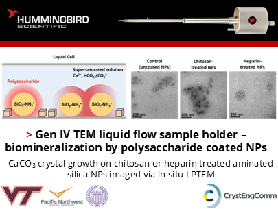
Brenna M. Knight, Patricia M. Dove, and their colleagues from Virginia Tech, PNNL, and University of Washington published research using the Hummingbird Scientific Gen IV liquid flow TEM sample holder to investigate how polysaccharide-functionalized nanoparticle interfaces influence CaCO₃ nucleation. Through direct in-situ liquid-phase TEM observation, the team showed that biomolecular interface chemistry plays an active role in controlling where crystallization begins, how rapidly nuclei emerge, and the spatial organization of resulting mineral formation. Their findings provide new mechanistic insight into biomineralization processes and the broader role of macromolecular interfacial environments in directing crystallization pathways.
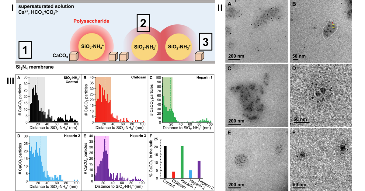
(I) Cross-sectional schematic of the in situ liquid TEM cell showing CaCO₃ nucleation near polysaccharide-coated silica nanoparticles. Highlighted interfaces: (1) Si₃N₄ membrane–solution, (2) polysaccharide–solution, and (3) polysaccharide–membrane–solution. Chitosan and heparin promote preferential nucleation at interface (3), producing ring-like CaCO₃ growth. (II) In situ LP-TEM images of CaCO₃ crystallites ~10 min after nucleation. (A,B) Control SiO₂–NH₃⁺ nanoparticles, (C,D) chitosan-treated, and (E,F) heparin-treated. Larger dark spheres are SiO₂ nanoparticles, smaller dark spots are CaCO₃, and the higher-density region around the nanoparticles is outlined in white. (III) Distance distributions from each CaCO₃ crystal to the nearest visible SiO₂–NH₃⁺ nanoparticle (up to 100 nm): (A) control, (B) chitosan-treated, and (C–E) three heparin-treated trials. Shaded regions indicate the electron-dense cloud, dotted lines show average cloud thickness, and (F) summarizes bulk nucleation frequency.
To capture these dynamic processes in real time, functionalized silica nanoparticles coated with either chitosan or highly charged heparin were encapsulated in a liquid cell with supersaturated calcium carbonate precursor solution and imaged under low electron dose conditions. The experiments revealed that heparin created a significantly stronger nucleation environment, driving earlier onset crystallization, higher nucleation density, and more spatially controlled crystal formation compared with chitosan-coated interfaces. By enabling direct visualization of transient nucleation events under realistic liquid conditions, the Hummingbird Scientific Gen IV liquid flow TEM sample holder provides a powerful platform for investigating crystallization, interfacial chemistry, biomineralization, and other dynamic solution-phase transformations that are inaccessible through conventional endpoint analysis.
Reference: Brenna M. Knight, Biao Jin, Yuna Bae, James J. De Yoreo, and Patricia M. Dove, CrystEngComm 28, 2835-2844 (2026) DOI: 10.1039/D6CE00012F
Full paper Copyright © 2026 The Authors. Published by Royal Society of Chemistry. This publication is licensed under CC BY-NC 3.0.
View All News