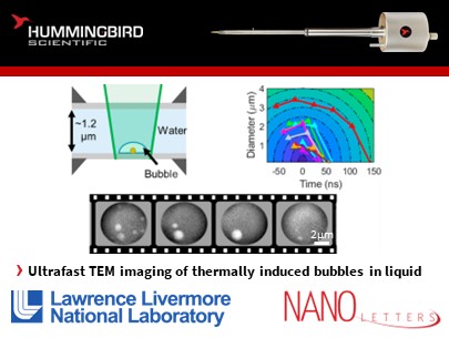
Researchers at Lawrence Livermore National Laboratory (LLNL) investigated the behavior of bubbles at the micron and nanoscale using ultrafast transmission electron microscopy to enhance our understanding of bubble dynamics in various applications such as microfluidics, drug delivery, and bubble-mediated therapies.
They used Hummingbird Scientific’s in-situ liquid cell TEM holder in the movie-mode dynamic transmission electron microscope at LLNL to capture with high spatial and temporal resolution the effect of laser heating metal nanoparticles in a liquid and the bubbles that formed at those particles.
Their findings revealed intricate bubble dynamics at the micron and nanoscale, shedding light on the fundamental mechanisms involved and providing valuable insights for designing more efficient bubble-based systems. Specifically, they showed that nanobubbles were observed to collapse quickly (50 ns), while micrometer scale bubbles were observed to first grow and then still collapse after a longer time (200 ns). These collapsing time scales were found to be consistent with modeling and data from other studies.
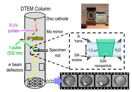
Illustration of their experimental setup: Laser pulses were used to generate electron pulses in the TEM electron source. The electron pulses were deflected onto different regions of the CCD. The CCD is then read, and the produced images arranged sequentially to produce a 9-frame time-sequence. Bubble formation inside the in-situ TEM liquid cell was initiated by a laser pulse with heat absorbing gold nanoparticle deposited onto the liquid cell. Copyright © 2022 American Chemical Society
Reference: Garth C. Egan, Edmond Y. Lau, and Eric Schwegler “Multiframe Imaging of Micron and Nanoscale Bubble Dynamics”, Nano Lett. 2022, 22, 3, 1053–1058. Copyright © 2022 American Chemical Society. Full Paper
View All News