How can electrodeposited nanoparticle growth and morphology be controlled?
Magdalena Parlinska-Wojtan, Tomasz Roman Tarnawski, See Wee Chee, and their colleagues from the Institute of Nuclear Physics Polish Academy of Sciences, Fritz-Haber-Institute of the Max-Planck Society, Biological and Chemical Research Centre, the Solaris National Synchrotron Radiation Centre at Jagiellonian University, Silesian University, and ETH Zürich published work using the Generation V Hummingbird Scientific In-situ Liquid Electrochemistry TEM sample holder to characterize the growth, morphology, and chemistry of electrodeposited PtNi nanoparticle films.

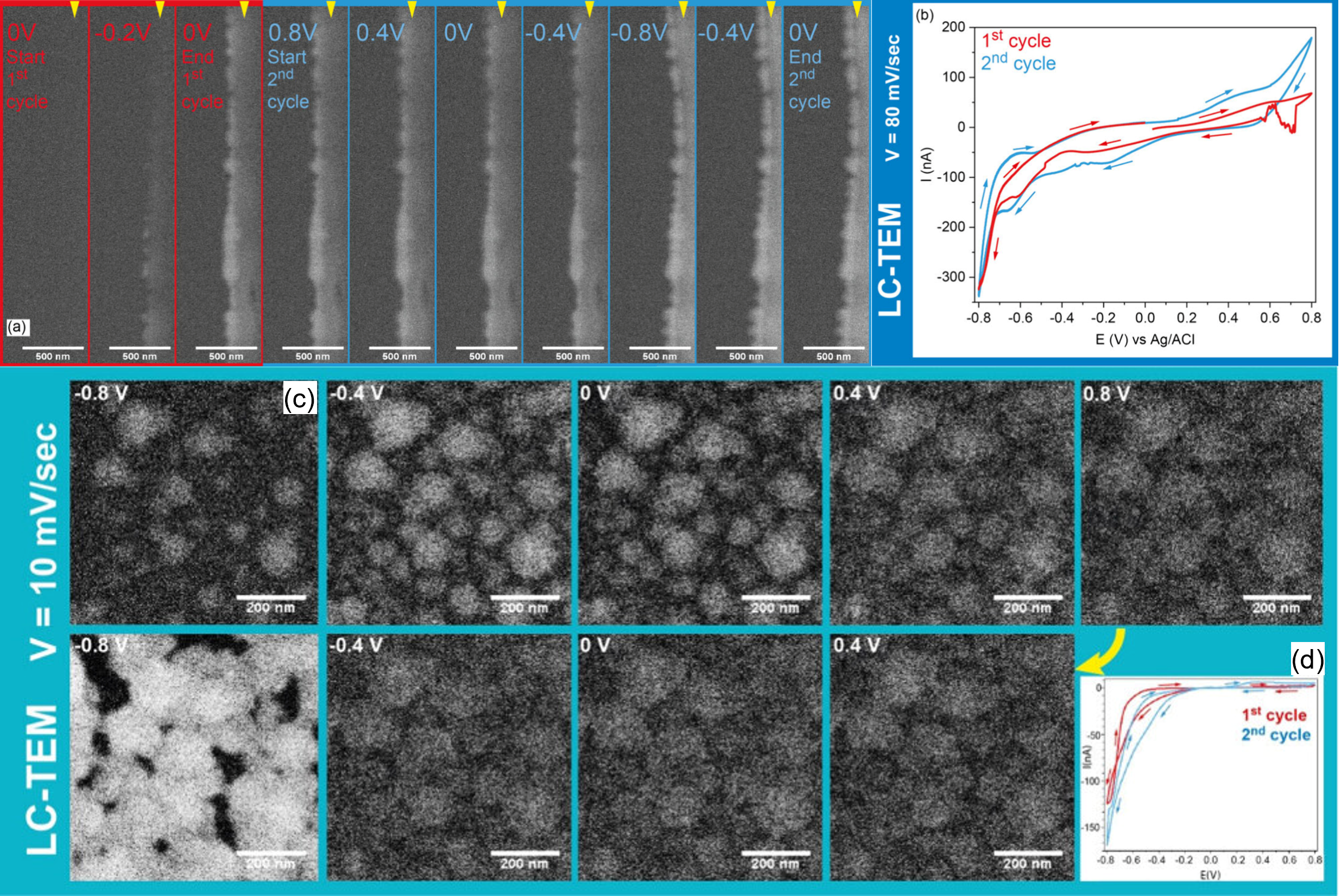
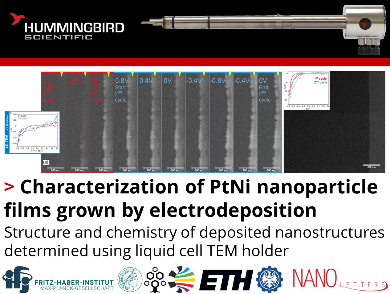
In-situ LC-TEM image sequences showing the multi-cycle electrodeposition of PtNi. a) Frames extracted at different applied potentials and b) current–potential curves obtained from scanning the potential between 0.8 and −0.8 V vs Ag/AgCl over with a scan rate of 80 mV/s. The Hummingbird Scientific holder comes with a customized Ag/AgCl reference electrode built into the holder which the applied potential is referenced to. The yellow triangles indicate the electrode edge, which is on the right side of the images. c) Frames corresponding to different applied potentials. d) Current vs potential curves of the first and second cycles of PtNi electrodeposition. Copyright © 2024 The Authors. Published by American Chemical Society. This publication is licensed under CC-BY 4.0
PtNi nanopartle films were grown on a carbon electrode using cyclic voltammetry of different scanning rates, which along with the number of cycles was found to be the most influential parameter on film thickness, homogeneity, nanoparticle size, and surface microstructure. Film thickness increases with continued cycling, leading to branched and porous structures. Precursor concentration did not significantly influence these features, but the applied potential range was found to influence the film chemistry, as measured by scanning transmission electron microscopy with energy dispersive X-ray spectroscopy (STEM-EDS) and scanning transmission X-ray microscopy (STXM), which confirmed that Ni was deposited in the oxide phase. The determination of these dominant parameters will inform future optimization of multicomponent nanoparticle film growth for energy and catalysis applications.
Reference:
Magdalena Parlinska-Wojtan, Tomasz Roman Tarnawski, Joanna Depciuch, Maria Letizia De Marco, Kamil Sobczak, Krzysztof Matlak, Mirosława Pawlyta, Robin E. Schaeublin, and See Wee Chee, Nano Letters XXXX (XXX) XXX-XXX (2024) DOI: 10.1021/acs.nanolett.4c02228
Full paper Copyright © 2024 The Authors. Published by American Chemical Society. This publication is licensed under CC-BY 4.0
View All News

