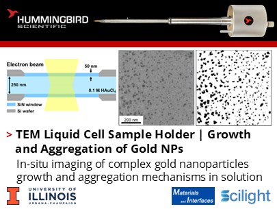
How do you model gold nanoparticle formation beyond classical theories of particle growth to capture coalescence, multistage complexity and growth?
Thao Ngo, Siying Yu, and Hong Yang from the Department of Chemical and Biomolecular Engineering, University of Illinois Urbana-Champaign used Hummingbird Scientific’s liquid-cell TEM sample holder to observe and quantify the dynamic formation of gold nanoparticles in solution. Unlike traditional ex situ methods, Liquid-cell TEM enables real-time visualization of nucleation, growth, and aggregation processes. The study developed a global analytical model combining diffusion, surface reaction, and coalescence to describe ensemble nanoparticle formation.
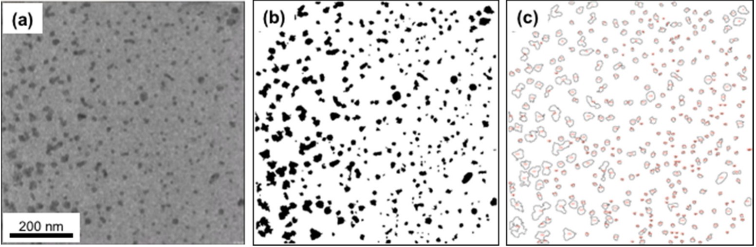
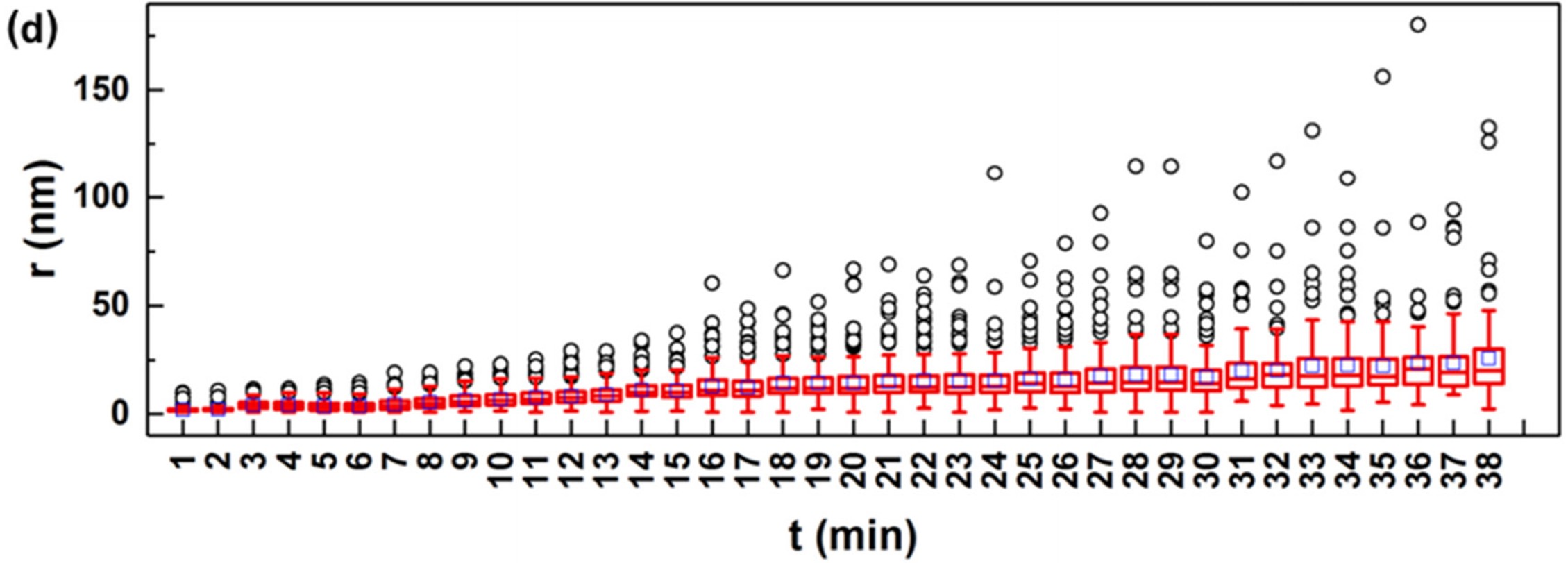
Illustration of the analysis of particle number and size from a LTEM micrograph using ImageJ software. (a) Still frame extracted from the LTEM video recorded during the growth, (b) processed image based on the contrast, and (c) image showing the outlines of the counted nanoparticles. (d) Particle size (r) over time (t) in the form of a box plot with outliers. The line and blue square inside each box represent the median and average particle size at that time, respectively. The top of the box represents the upper quartile while the bottom of the box represents the lower quartile. The upper and lower whiskers represent particles with sizes that fall outside the middle 50%. Copyright © 2025 Scilight
The study quantitatively analyzed gold nanoparticle formation and evaluated classical Lifshitz–Slyozov–Wagner (LSW) theory alongside coagulation kinetics. Since neither model fully explained the process, a polynomial expression was developed to capture diffusion and reaction controlled growth combined with particle coalescence. This approach describes the complexity across three stages: initial monomer addition (Stage I), growth with coalescence (Stage II), and dendritic formation (Stage III). While the polynomial fit aids simulation of nanoparticle formation in solution, fully understanding the underlying mechanisms remains challenging.
This approach offers a framework for simulating nanoparticle formation under realistic conditions, enabling better control of size and morphology for applications in catalysis, biomedicine, and nanotechnology.
Reference: Thao Ngo, Siying Yu, and Hong Yang, H. Mater. Interfaces 2025, 2(2), 201–212. DOI: 10.53941/mi.2025.100016
Full paper Copyright © 2025 Scilight, Mater. Interfaces. All rights are reserved
View All News