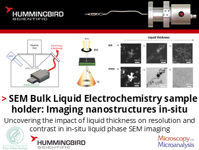
How does the SEM bulk liquid electrochemistry holder enable high-resolution imaging of nanomaterials in liquid environments within an SEM?
Aram Yoon, See Wee Chee, Beatriz Roldán Cuenya, and their colleagues at the Fritz-Haber-Institute of the Max-Planck Society in collaboration with Hummingbird Scientific published on their use of the Hummingbird Scientific in-situ SEM bulk liquid electrochemistry sample holder (and demonstrated the capability to image nanostructures in liquid electrolyte environment at high contrast and in high resolution down to tens of nanometers in real-time.
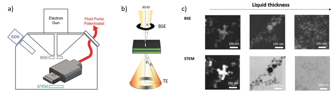
(a) Diagram of the electrochemical LC-SEM system featuring BSE, STEM, and EDX detectors. Fluid and electrical connections are routed through tubing and wiring linked to an external syringe pump and potentiostat. (b) Scattered electrons from the liquid cell are detected in both forward (STEM) and backward (BSE) directions. The electron beam has a convergence angle (α) of 2 mrad, with the STEM detector collecting electrons over a 0–250 mrad range (β). (c) Simultaneously acquired BSE and STEM images of Cu₂O nanoparticles show changes with increasing liquid layer thickness, progressing from left to right. In the STEM images, contrast inversion becomes apparent as the liquid layer thickens.
The study demonstrated the capability to image nanostructures in liquid electrolyte environment at high contrast and in high resolution down to tens of nanometers in-situ. Copper oxide nanocubes were investigated in different thickness of electrolyte layers in two different imaging modes – backscattered (BSE) and transmitted (TE) electrons. Images obtained using BSE mode retain high spatial resolution but suffer from decreasing signal-to-noise ratios with increasing liquid thickness. Likewise, images acquired using TEs show better contrast with some sacrifice in the spatial resolution (Figure below). The Hummingbird Scientific in-situ SEM bulk liquid electrochemistry sample holder that incorporates real reference electrode (Ag/AgCl) and real counter electrode (Pt) provides a powerful liquid cell SEM platform to measure and observe realistic electrochemistry with accurate quantitative information.
Reference: Aram Yoon, Antonia Herzog, Philipp Grosse, Daan Hein Alsem, See Wee Chee and Beatriz Roldán Cuenya, Microscopy and Microanalysis 27, 1 (2021). DOI: 10.1017/S1431927620024769
Full paper From Microscopy and Microanalysis under the Creative Commons license
View All News