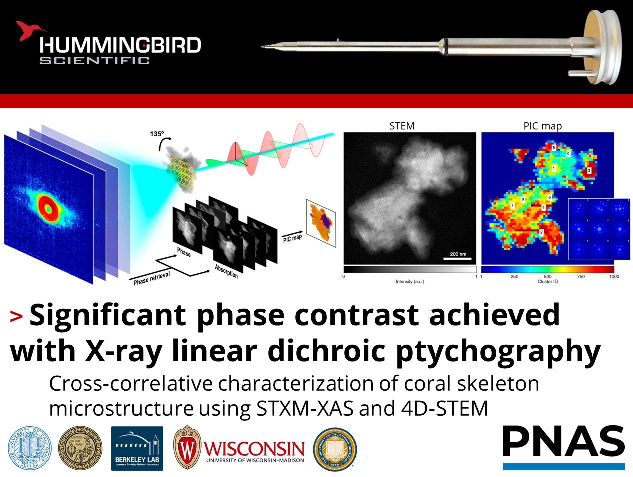
Curious about the microstructure of coral skeletons?
Yuan Hung Lo, Jianwei Miao, and their colleagues at the University of California- Los Angeles and University of Colorado- Boulder published work using the Hummingbird Scientific tomography sample holder to perform cross-correlative X-ray linear dichroic ptychography and 4D scanning transmission electron microscopy (4D-STEM) and tomography on coral skeletons. The orientation microstructure of the calcium carbonate nanocrystals were mounted in the Hummingbird Scientific tomography holder for electron microscopy and to collect ptychographic data at multiple X-ray polarizations and energies around the oxygen K-edge.
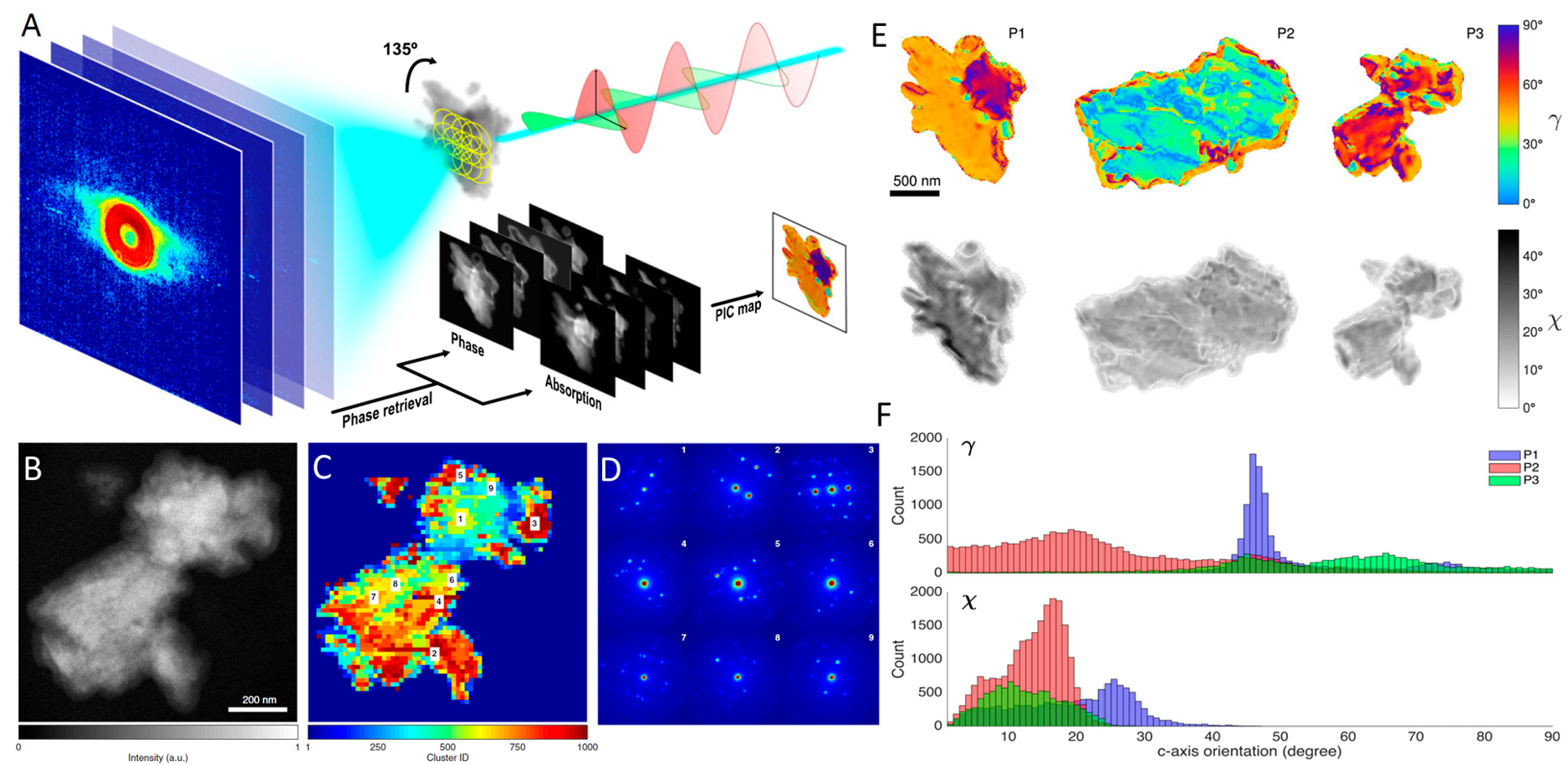
a) Horizontally and vertically polarized X-rays incident on the specimen as spatially overlapping diffraction patterns were acquired below (534.5 eV) and on (536.5 eV) the O K-edge absorption edge to obtain polarization data at various rotation angles (left). Experimental schematic of X-ray linear dichroic ptychography (right). b) STEM image of nanoparticle used to acquire scanning electron nano-diffraction patterns. c) Crystal axis similarity map generated using hierarchical clustering of diffraction patterns. Comparable color indicates subdomains with similar crystal orientations. d) Representative CBED patterns from various regions of the coral nanoparticle showing nanoscale orientational diversity. e) Quantitative PIC maps of the three aragonite particles, calculated using 0°, 45°, and 90° linear dichroic ptychography images. The color map denotes in-plane azimuthal crystal c-axis angle (γ), while the greyscale denotes out-of-plane c-axis angle (χ). f) Histograms of in-plane (γ) and out-of-plane (χ) angles for the three particles, suggesting the presence of both spherulitic and randomly oriented crystallites. Copyright 2021 National Academy of Sciences
The ptychography phase and absorption images exhibited striking polarization dependence, revealing the presence of differently oriented nanodomains across the coral fragments. Quantitative polarization-based analysis uncovered surprising orientational diversity, including both narrowly distributed and widely spread crystalline orientations down to 35nm resolution. Nanoscale crystallographic trends were confirmed using 4D-STEM. The electron diffraction analyses qualitatively corroborated the X-ray results, confirming the presence of disconnected yet co-oriented crystallites. This correlative workflow combining X-ray and electron microscopy functionalities with consistent sample delivery hardware enabled new insights into the structure and formation of biominerals.
Reference: Yuan Hung Lo, Jihan Zhou, Arjun Rana, Drew Morrill, Christian Gentry, Bjoern Enders, Young-Sang Yu, Chang-Yu Sun, David A. Shapiro, Roger W. Falcone, Henry C. Kapteyn, Margaret M. Murnane, Pupa U. P. A. Gilbert and Jianwei Miao, Proc. Natl. Acad. Sci. 118 (3) e2019068118 (2021) DOI: 10.1073/pnas.2019068118
Full paper Copyright © 2021 National Academy of Sciences
View All News