Curious about how corrosion initiates in iron at the nanoscale?
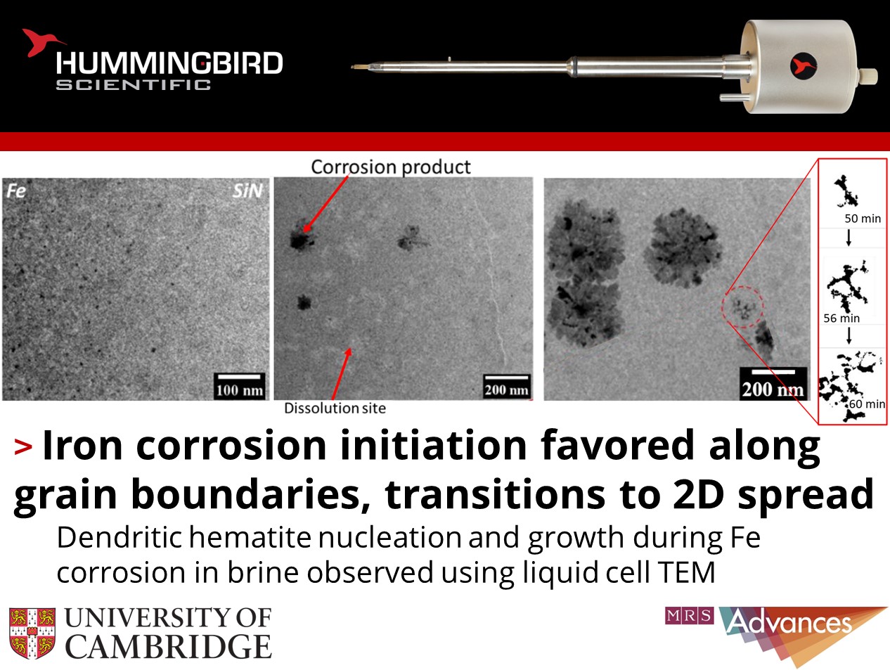
Surabhi Agrawal, Stuart M. Clarke and their colleagues at the University of Cambridge published in-situ observation of iron corrosion in brine using their Hummingbird Scientific in-situ liquid cell TEM sample holder. Inside of the liquid cell, dissolution of a 50 nm iron film and the nucleation and growth of hematite corrosion products is captured during exposure to flowing brine.
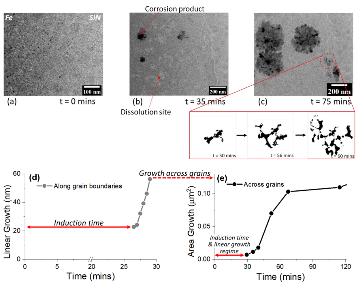
Figures showing a) uncorroded iron film at beginning of experiment, b) after 35 minutes, three corrosion product nuclei have appeared, c) after 75 minutes these sites grow and new sites nucleate, insert with processed binary images showing shape evolution. d) Plot of slow linear growth and incubation in first 25 minutes of the experiment. e) Plot of more rapid areal growth across grain surfaces. Copyright © 2023 Springer Nature
Dendritic hematite nuclei emerged on the metal surface preferentially along grain boundaries. This evidences grain boundaries as critical active sites for nucleation of dissolved Fe. Quantitative image analysis revealed an initial linear growth regime spreading up to 3nm/s along boundaries, transitioning to a 2D growth regime exceeding growth rates of 50nm2/s. In-situ selected area electron diffraction patterns taken at localized corrosion regions confirmed the iron oxide phase as hematite. This analytical capability highlights the power of liquid cell TEM to correlate real-time observations with microstructural data. Further characterization of such ephemeral reaction dynamics will inform predictive models and corrosion mitigation strategies.
Reference: Surabhi Agrawal, Mobbassar H. Sk, Richard M. Langford, Stuart M. Clarke, MRS Advances 8 376−380 (2023) DOI: 10.1557/s43580-023-00533-1
Full paper Copyright © 2023 Springer Nature
View All News