Researchers led by Tanya Prozorov et al. at Ames National Laboratory have published a new method to image gel-based nanocomposites using a liquid cell TEM, which is traditionally performed using cryo-based TEM technique. The work performed using the Hummingbird Scientific’s liquid cell platform opens a new avenue to image particles in thick gel media devoid of any artifacts. The details have been published in the Journal Micron.
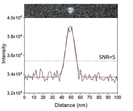
Traditionally, imaging polymeric particles in their thick gel layers have been limited to cryo-TEM vitreous imaging technique, which is notorious for tedious sample preparation steps and introducing artifacts during the process. This limitation inspired the researchers at Ames to develop a new method to image gel-based nanocomposites in their native environment using the liquid cell holder developed by Hummingbird Scientific. The researchers used the controlled deposition of femtoliter-range viscous specimen into the SiN windows of the liquid cell assembly and imaged particles as small as 5 nm dispersed in 600 nm thick gel layer (image above). The method developed here can pave the way for imaging particles while in their fluidic environment with free of artifacts and less e-beam damage.
Reference: Alejandra Londono-Calderon, Srikanth Nayak, Curtis L. Mosher, Surya K. Mallapragada, and Tanya Prozorov. “New Approach to Electron Microscopy Imaging of Gel Nanocomposites in situ,” Micron (2019). DOI: 10.1016/j.micron.2019.02.010
View All News